Confocal Microscopy

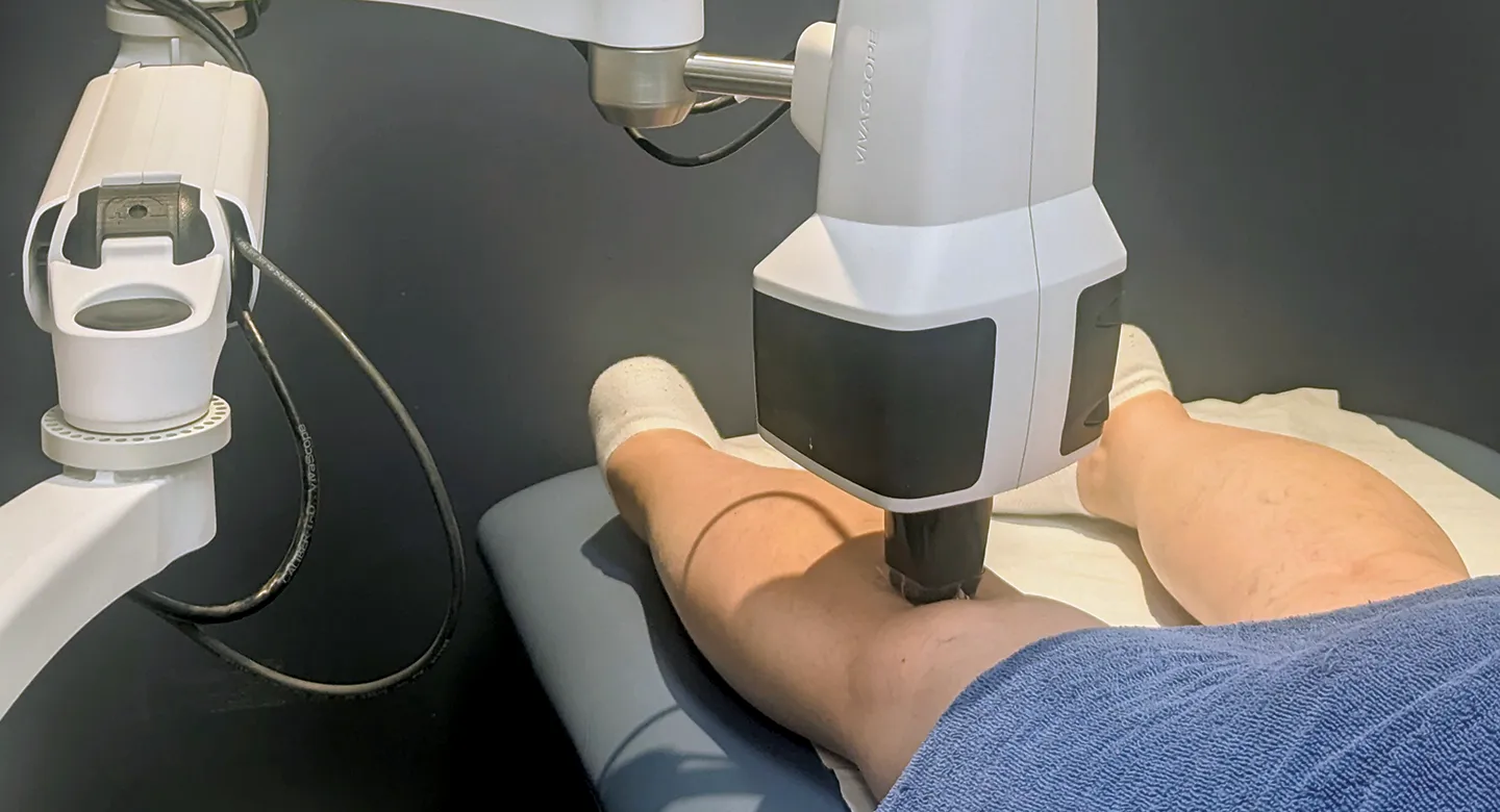
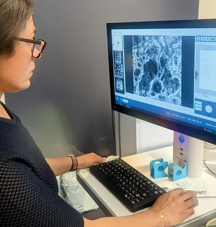
Confocal microscopy allows doctors to examine individual skin cells in real time without needing to remove tissue. Using a laser microscope, a confocal microscopy provides magnification of up to 1000 times, offering a cellular-level view of the upper layers of the skin.
Confocal microscopy is especially prized for its high degree of accuracy in diagnosing skin cancer, including melanoma. It allows clinicians to distinguish between benign (non-cancerous) and malignant lesions, reducing the need for unnecessary biopsies. Studies have shown that confocal microscopy can lower biopsy rates by up to 43% while maintaining a melanoma detection sensitivity of 92%.
Confocal microscopy is also useful in treatment planning and follow-up. It can guide where to biopsy within a suspicious lesion and help map the edges of a melanoma before surgery. This is especially valuable in challenging cases such as lentigo maligna on the face. Additionally, confocal microscopy can monitor how well non-surgical treatments (including topical therapies and/or radiotherapy) are working.


As a subspecialty of dermatology, confocal microscopy requires advanced training and deep knowledge of skin pathology to interpret the highly detailed images effectively. At Austin Clinic, Dr Mehrnoosh Alian provides exactly that. An experienced general practitioner, she completed her medical degree at Tabriz University of Medical Sciences, graduating with a Doctorate in Medicine. She has since undertaken extensive post-graduate education in skin cancer detection and management.
Book your appointment with Dr Alian here.